Rat Anti-Mouse CD8α-AF488 (53-6.7)
Cat. No.:
1550-30
Alexa Fluor® 488 Anti-Mouse CD8α antibody for use in flow cytometry and immunohistochemistry / immunocytochemistry assays.
$230.00


| Clone | 53-6.7 |
|---|---|
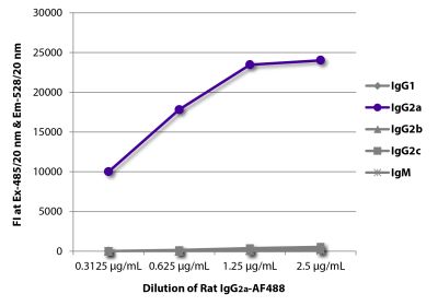
| Isotype | Rat (LOU/Ws1/M) IgG2aκ |
| Isotype Control | Rat IgG2a-AF488 (KLH/G2a-1-1) |
| Specificity | Mouse CD8α |
| Alternative Names | Ly-2, Lyt-2, Ly-35, Ly-B, T8 |
| Description | In the mouse, CD8 exists in two forms – (i) a CD8 heterodimer composed of an α chain (CD8α) and a β chain (CD8β); and (ii) a homodimer of two α chains. The heterodimer is found on the surface of essentially all thymocytes and the “suppressor/cytotoxic” subpopulation of mature T lymphocytes. Subsets of intestinal intraepithelial lymphocytes express CD8α without CD8β. It has been suggested that CD8+β- T cells mature extrathymically, while development of the CD8α+β+ population of T cells is thymus-dependent. CD8 acts as a coreceptor with MHC Class I-restricted T cell receptors in antigen recognition and positive selection of MHC class I-restricted CD8+ T cells. In vivo and in vitro treatment with the 53-6.7 monoclonal antibody effectively depletes CD8α+ cells. The 53-6.7 monoclonal antibody also blocks allogeneic help specific for class I MHC antigens and T cell responses to IL-2. |
| Immunogen | Spleen cells or thymocyte membranes |
| Conjugate | AF488 (Alexa Fluor® 488) |
| Buffer Formulation | Phosphate buffered saline containing < 0.1% sodium azide |
| Clonality | Monoclonal |
| Concentration | 0.5 mg/mL |
| Volume | 0.2 mL |
| Recommended Storage | 2-8°C; Avoid exposure to light |
| Trademark Information | Alexa Fluor® is a registered trademark of Thermo Fisher Scientific, Inc. or its subsidiaries |
| Applications |
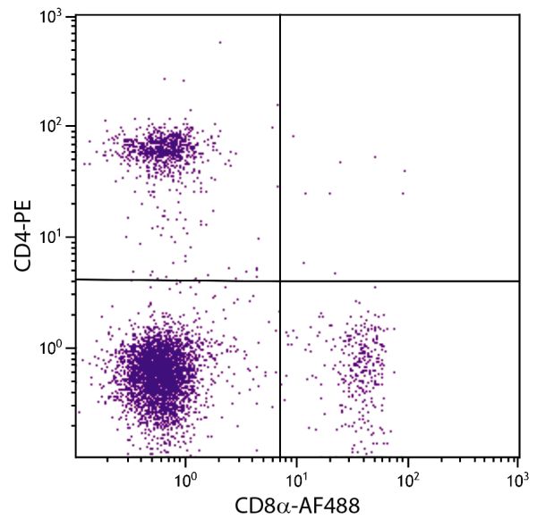
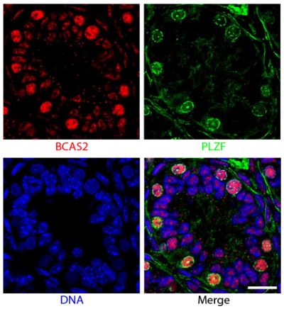
Flow Cytometry – Quality tested 1,6,7,9-15 Immunohistochemistry-Frozen Sections – Reported in literature 2-5 Immunocytochemistry – Reported in literature 7 Immunoprecipitation – Reported in literature 1,6 Depletion – Reported in literature 8 Blocking – Reported in literature 6 |
| RRID Number | AB_2794887 |
| Gene ID |
12525 (Mouse) |
| Gene ID Symbol |
Cd8a (Mouse) |
| Gene ID Aliases | Ly-2; Ly-B; Ly-35; Lyt-2; BB154331 |
| UniProt ID |
P01731 (Mouse |
| UniProt Name |
CD8A_MOUSE (Mouse) |
Documentation
Certificate of Analysis Lookup
Enter the Catalog Number and Lot Number for the Certificate of Analysis you wish to view
- 1. Ledbetter JA, Herzenberg LA. Xenogeneic monoclonal antibodies to mouse lymphoid differentiation antigens. Immunol Rev. 1979;47:63-90. (Immunogen, IP, FC)
- 2. Konno A, Takada K, Saegusa J, Takiguchi M. Presence of B7-2+ dendritic cells and expression of Th1 cytokines in the early development of sialodacryoadenitis in the IQI/Jic mouse model of primary Sjörgren's syndrome. Autoimmunity. 2003;36:247-54. (IHC-FS)
- 3. Delong P, Tanaka T, Kruklitis R, Henry AC, Kapoor V, Kaiser LR, et al. Use of cyclooxygenase-2 inhibition to enhance the efficacy of immunotherapy. Cancer Res. 2003;63:7845-52. (IHC-FS)
- 4. Wehling-Henricks M, Jordan MC, Gotoh T, Grody WW, Roos KP, Tidball JG. Arginine metabolism by macrophages promotes cardiac and muscle fibrosis in mdx muscular dystrophy. PloS One. 2010;5(5):e10763. (IHC-FS)
- 5. Kunikata N, Sano K, Honda M, Ishii K, Matsunaga J, Okuyama R, et al. Peritumoral CpG oligodeoxynucleotide treatment inhibits tumor growth and metastasis of B16F10 melanoma cells. J Invest Dermatol. 2004;123:395-402. (IHC-FS)
- 6. Ledbetter JA, Seaman WE, Tsu TT, Herzenberg LA. Lyt-2 and lyt-3 antigens are on two different polypeptide subunits linked by disulfide bonds. Relationship of subunits to T cell cytolytic activity. J Exp Med. 1981;153:1503-16. (IP, FC, Block)
- 7. Choi J, Oh S, Lee D, Oh HJ, Park JY, Lee SB, et al. Mst1-FoxO signaling protects naïve T lymphocytes from cellular oxidative stress in mice. PloS One. 2009;4(11):e8011. (FC, ICC)
- 8. Hathcock KS. T cell depletion by cytotoxic elimination. Curr Protoc Immunol. 1991;3:4.1. (Depletion)
- 9. Lagrota-Candido J, Vasconcellos R, Cavalcanti M, Bozza M, Savino W, Quirico-Santos T. Resolution of skeletal muscle inflammation in mdx dystrophic mouse is accompanied by increased immunoglobulin and interferon-γ production. Int J Exp Path. 2002;83:121-32. (FC)
- 10. Parmo-Cabañas M, García-Bernal D, García-Verdugo R, Kremer L, Márquez G, Teixidó J. Intracellular signaling required for CCL25-stimulated T cell adhesion mediated by the integrin α4β1. J Leukoc Biol. 2007;82:380-91. (FC)
- 11. Fink LN, Frøkiær H. Dendritic cells from Peyer's patches and mesenteric lymph nodes differ from spleen dendritic cells in their response to commensal gut bacteria. Scand J Immunol. 2008;68:270-9. (FC)
- 12. Jordan KR, Buhrman JD, Sprague J, Moore BL, Gao D, Kappler JW, et al. TCR hypervariable regions expressed by T cells that respond to effective tumor vaccines. Cancer Immunol Immunother. 2012;61:1627-38. (FC)
- 13. Elgbratt K, Jansson A, Hultgren-Hörnquist E. A quantitative study of the mechanisms behind thymic atrophy in Gαi2-deficient mice during colitis development. PLoS One. 2012;7(5):e36726. (FC)
- 14. Domingos-Pereira S, Decrausaz L, Derré L, Bobst M, Romero P, Schiller JT, et al. Intravaginal TLR agonists increase local vaccine-specific CD8 T cells and human papillomavirus-associated genital-tumor regression in mice. Mucosal Immunol. 2013;6:393-404. (FC)
- 15. Grodeland G, Mjaaland S, Roux KH, Fredriksen AB, Bogen B. DNA vaccine that targets hemagglutinin to MHC class II molecules rapidly induces antibody-mediated protection against influenza. J Immunol. 2013;191:3221-31. (FC)
See All References


