Mouse Anti-Human CD4-FITC (RFT4)
Only %1 left
Cat. No.:
9522-02,
9522-02S
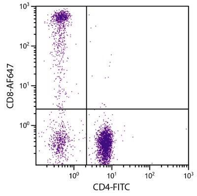
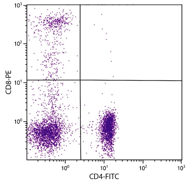
FITC Anti-Human CD4 antibody for use in flow cytometry, immunohistochemistry / immunocytochemistry, and western blot assays.
As low as
$50.00

| Clone | RFT4 |
|---|---|
| Isotype | Mouse (BALB/c) IgG1κ |
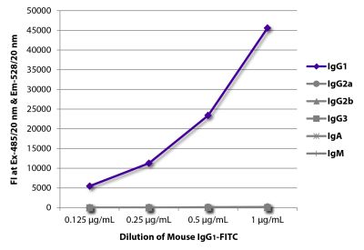
| Isotype Control | Mouse IgG1-FITC (15H6) |
| Specificity | Human CD4 |
| Alternative Names | T4, Leu-3 |
| Description | CD4 is a 55 kDa type I transmembrane glycoprotein and a member of the Ig-SF of cell surface receptors. It is expressed predominantly on the “helper/inducer” subpopulation of mature T lymphocytes and on monocytes and macrophages. Domains 1 and 2 bind to MHC Class II antigens, while domains 3 and 4 may be involved in cis interactions with the CD3/TCR complex. CD4 functions as an accessory molecule in the recognition of foreign antigens in association with MHC class II antigens by T cells. |
| Immunogen | Thymocytes and E-rosetted lymphocytes |
| Conjugate | FITC (Fluorescein) |
| Buffer Formulation | Phosphate buffered saline containing < 0.1% sodium azide |
| Clonality | Monoclonal |
| Concentration | Lot specific |
| Volume | 0.25 mL or 1.0 mL |
| Recommended Storage | 2-8°C; Avoid exposure to light |
| Applications |
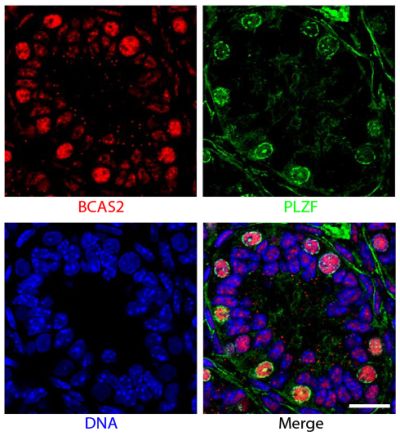
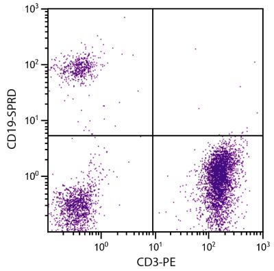
Flow Cytometry – Quality tested 1,7-9 Immunohistochemistry-Frozen Sections – Reported in literature 3 Immunocytochemistry – Reported in literature 4,5 Western Blot – Reported in literature 2 Separation – Reported in literature 6 |
| RRID Number | AB_2796846 |
| Gene ID |
920 (Human) |
| Gene ID Symbol |
CD4 (Human) |
| Gene ID Aliases | CD4mut |
| UniProt ID |
P01730 (Human) |
| UniProt Name |
CD4_HUMAN (Human) |
Documentation
Certificate of Analysis Lookup
Enter the Catalog Number and Lot Number for the Certificate of Analysis you wish to view
- 1. Sattentau QJ, Arthos J, Deen K, Hanna N, Healey D, Beverley PC, et al. Structural analysis of the human immunodeficiency virus-binding domain of CD4. Epitope mapping with site-directed mutants and anti-idiotypes. J Exp Med. 1989;170:1319-34. (FC)
- 2. Eugene HS, Pierce-Paul BR, Craigo JK, Ross TM. Rhesus macaques vaccinated with consensus envelopes elicit partially protective immune responses against SHIV SF162p4 challenge. Virol J. 2013;10:102. (WB)
- 3. Hufert FT, van Lunzen J, Janossy G, Bertram S, Schmitz J, Haller O, et al. Germinal centre CD4+ T cells are an important site of HIV replication in vivo. AIDS. 1997;11:849-57. (IHC-FS)
- 4. Poulter LW, Norris A, Powers C, Condez A, Burnes H, Schmekel B, et al. T cell dominated inflammatory reactions in the bronchioles of asymptomatic asthmatics are also present in the nasal mucosa. Postgrad Med J. 1991;67:747-53. (ICC)
- 5. Burgess R, Hyde K, Maguire PJ, Kelsey PR, Yin JA, Geary CG. Two-colour immunoenzymatic technique using sequential staining by APAAP to evaluate two cell antigens. J Clin Pathol. 1992;45:206-9. (ICC)
- 6. D'Acquisto F, Paschalidis N, Raza K, Buckley CD, Flower RJ, Perretti M. Glucocorticoid treatment inhibits annexin-1 expression in rheumatoid arthritis CD4+ T cells. Rheumatology. 2008;47:636-9. (Sep)
- 7. Daubenberger CA, Salomon M, Vecino W, Hübner B, Troll H, Rodriques R, et al. Functional and structural similarity of Vγ9V δ2 T cells in humans and Aotus monkeys, a primate infection model for Plasmodium falciparum malaria. J Immunol. 2001;167:6421-30. (FC)
- 8. Zariffard MR, Saifuddin M, Finnegan A, Spear GT. HSV type 2 infection increases HIV DNA detection in vaginal tissue of mice expressing human CD4 and CCR5. AIDS Res Hum Retroviruses. 2009;25:1157-64. (FC)
- 9. Roetynck S, Olotu A, Simam J, Marsh K, Stockinger B, Urban B, et al. Phenotypic and functional profiling of CD4 T cell compartment in distinct populations of healthy adults with different antigenic exposure. PLoS One. 2013;8(1):e55195. (FC)
See More


