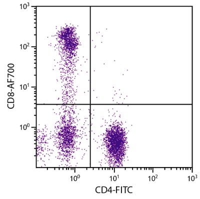
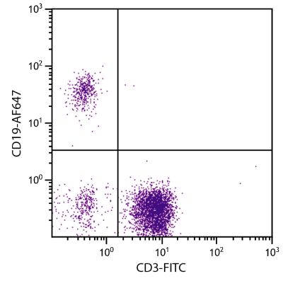
Mouse Anti-Human CD16-BIOT (GRM1)
Cat. No.:
9570-08
Biotin Anti-Human CD16 antibody for use in flow cytometry, immunohistochemistry, western blot, immunoprecipitation, and ELISA assays.
$206.00
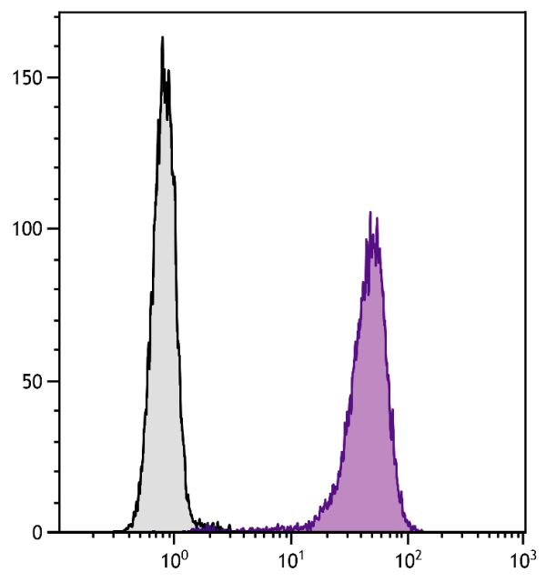

| Clone | GRM1 |
|---|---|
| Isotype | Mouse (BALB/c) IgG2aκ |
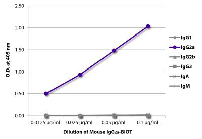
| Isotype Control | Mouse IgG2a-BIOT (HOPC-1) |
| Specificity | Human CD16 |
| Alternative Names | FcγRIII, low affinity Fc receptor |
| Description | CD16, a member of the immunoglobulin superfamily, is a 50-65 kDa glycoprotein found as both a transmembrane and GPI-linked form. The transmembrane form of CD16 is expressed on NK cells, granulocytes, macrophages, and mast cells but not on eosinophils. The GPI-anchored type of CD16 is found only on neutrophils. CD16 is involved in NK activation and signal transduction. |
| Immunogen | Mononuclear cells from a prolymphocytic B-leukemia |
| Conjugate | BIOT (Biotin) |
| Buffer Formulation | Phosphate buffered saline containing < 0.1% sodium azide |
| Clonality | Monoclonal |
| Concentration | Lot specific |
| Volume | 1.0 mL |
| Recommended Storage | 2-8°C |
| Applications |
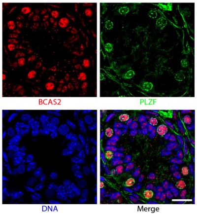
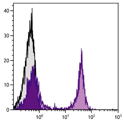
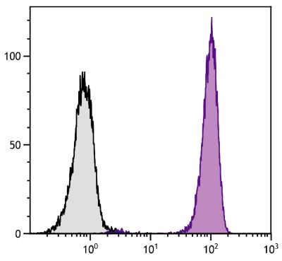
Flow Cytometry – Quality tested 6 Immunohistochemistry-Frozen Sections – Reported in literature 3 Immunoprecipitation – Reported in literature 1,2 Western Blot – Reported in literature 3 ELISA – Reported in literature 4,5 Complement Mediated Cell Depletion – Reported in literature 1 |
| RRID Number | AB_2796955 |
| Gene ID |
2214 (Human) |
| Gene ID Symbol |
FCGR3A (Human) |
| Gene ID Aliases | CD16; CD16A; FCG3; FCGR3; FCGRIII; FCR-10; FCRIII; FCRIIIA; IGFR3; IMD20 |
| UniProt ID |
P08637 (Human |
| UniProt Name |
FCG3A_HUMAN (Human) |
Documentation
Certificate of Analysis Lookup
Enter the Catalog Number and Lot Number for the Certificate of Analysis you wish to view
- 1. Lopez-Nevot MA, Cabrera T, Huelin C, Ruiz-Cabello F, Garrido F. A study of mAb GRM1 which reacts with natural killer cells and granulocytes. In: McMichael AJ, Beverley PC, Cobbold S, Crumpton MJ, Gilks W, Gotch FM, et al, editors. Leukocyte Typing III: White Cell Differentiation Antigens. Oxford: Oxford University Press; 1987. p. 707. (Immunogen, IP, CMDC)
- 2. Uggla CK, Jondal M, Kaplan D, Flomenberg N, Knowles RW. Enhancement of natural killer cell activity by unique antibodies within the CD2 (sheep-RBC receptor) and CD16 (Fc receptor) clusters. In: McMichael AJ, Beverley PC, Cobbold S, Crumpton MJ, Gilks W, Gotch FM, et al, editors. Leukocyte Typing III: White Cell Differentiation Antigens. Oxford: Oxford University Press; 1987. p. 134-7. (IP)
- 3. Wainwright SD, Holmes CH. Distribution of Fcγ receptors on trophoblast during human placental development: an immunohistochemical and immunoblotting study. Immunology. 1993;80:343-51. (IHC-FS, WB)
- 4. Masuda M, Miyoshi H, Kobatake S, Nishimura N, Dong XH, Komiyama Y, et al. Increased soluble FcγRIIIaMφ in plasma from patients with coronary artery diseases. Artherosclerosis. 2006;188:377-83. (ELISA)
- 5. Masuda M, Amano K, Hong SY, Nishimura N, Fukui M, Yoshika M, et al. Soluble FcγRIIIaMφ levels in plasma correlate with carotid maximum intima-media thickness (IMT) in subjects undergoing an annual medical checkup. Mol Med. 2008;14:436-42. (ELISA)
- 6. Pilling D, Vakil V, Gomer RH. Improved serum-free culture conditions for the differentiation of human and murine fibrocytes. J Immunol Methods. 2009;351:62-70. (FC)
See All References


