Share a link
Introduction to Western Blotting
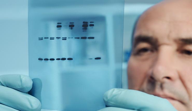
Western blot is a technique that involves separating a complex mixture of proteins by polyacrylamide gel electrophoresis (PAGE) before transferring them to a membrane for antibody-based detection. Advantages of Western blot are that it is relatively simple to perform and provides easily interpretable results, making it one of the most popular immunoassay formats available.
A typical Western blot workflow involves a series of steps, beginning with sample preparation. Next, gel electrophoresis and protein transfer are performed, followed by target detection with suitable antibody reagents. After washing to remove any unbound antibodies, the workflow concludes with image acquisition and analysis to provide either qualitative or quantitative data depending on the chosen method. Each of the Western blot steps must be carefully optimized to ensure accurate, reproducible results.

Western Blot Sample Preparation
Cell lysates and tissue homogenates are the most common sample types analyzed by Western blot. To prepare cell lysates, the cells are washed to remove any growth media components or cellular debris and are then incubated in an ice-cold lysis buffer matched to the target of interest. While buffers based on NP-40 or Triton X-100 are often used for preparing whole cell lysates or cytoplasmic extracts, more stringent lysis conditions (e.g., RIPA buffer) are typically required for extracting nuclear or mitochondrial proteins. Next, the lysates are cleared by centrifugation and the supernatants are analyzed. Tissue homogenates are prepared in a similar way, but the samples must first be dissociated using mechanical or enzymatic methods.
Electrophoretic Separation of Proteins
Following sample preparation, the proteins are separated based on their size and/or charge. This is achieved by loading the samples onto a gel and applying an electrical current, which causes the proteins to migrate from the negatively charged cathode at the top of the gel toward the positively charged anode at the bottom. Almost all Western blot gels are made of polyacrylamide, which serves as a molecular sieve, hence the term polyacrylamide electrophoresis (PAGE).
Two main types of PAGE are used for Western blotting. The first of these, known as native or non-denaturing PAGE, preserves the native protein structure to enable separation based on the mass: charge ratio. The second, sodium dodecyl sulfate-PAGE (SDS-PAGE), separates proteins according to size alone as the SDS confers a strong negative charge to each protein. In addition, SDS-PAGE uses a reducing agent such as dithiothreitol (DTT) or beta-mercaptoethanol (BME) to disrupt disulfide bonds and reduce proteins to their primary structure.
Although SDS-PAGE is more common, native gels are widely used for studying protein complexes or for situations where enzymatic activity must be maintained.
-
Sample Normalization for Western Blot
Normalization serves to allow a direct comparison between different samples on the same Western blot. It involves determining the total protein concentration of each sample (e.g., by performing a Bradford assay or using UV spectrophotometry) before diluting all of the samples to the same concentration with lysis buffer. Sample loading buffer is then added, which varies in composition depending on whether native or denaturing conditions are required.
-
Choosing the right polyacrylamide gel for Western blotting
Polyacrylamide gel selection is largely based on the size of the target protein. Generally, higher percentage polyacrylamide gels should be used for low molecular weight proteins, and vice versa, although gradient gels are often preferred for resolving similar-sized proteins or studying a broad range of protein sizes simultaneously. Pairing the gel to an appropriate running buffer is key to optimal separation; for example, MES buffer is often used for separating low molecular weight proteins while MOPS is more suitable for separating larger proteins. Critically, running buffers used for native Western blotting should not contain SDS. Gel manufacturers can usually offer guidance for product selection.
Protein transfer
Following gel electrophoresis, the separated proteins must be transferred onto a membrane to allow for antibody-based detection. The most common method, electrophoretic transfer, involves placing the gel in direct contact with the membrane (ensuring no air bubbles are present) and sandwiching the gel-membrane pairing between filter papers and porous sponge pads to facilitate the transfer process. The resultant sandwich is then placed between two electrodes in a conducting solution (the transfer buffer). When an electrical field is applied, the proteins are driven out of the gel and onto the membrane, where they become tightly bound.
-
Wet transfer vs. semi-dry transfer
During a wet transfer, the sandwich is fully submerged in the transfer buffer. This method provides a high transfer efficiency for a broad range of protein sizes, but takes hours to overnight. A semi-dry transfer instead requires only that the sandwich be saturated in the transfer buffer and usually takes less than an hour. However, semi-dry transfer is unsuitable for larger proteins (>300 kDa), which may remain trapped in the gel. An alternative approach, known as a dry transfer, uses no buffer at all and takes only minutes to transfer both small and large proteins, but comes with higher associated costs.
-
Western blot membrane types
Nitrocellulose and polyvinylidene fluoride (PVDF) are the two main membrane types used for Western blotting. While nitrocellulose benefits from an extremely high protein binding capacity, it can be brittle, limiting its utility for repeated stripping and reprobing. The superior strength of PVDF means it is often used where further protein characterization will be performed (e.g., sequencing applications), yet PVDF can sometimes produce higher background staining. Membrane selection is largely down to personal preference and the aims of the study.
-
Blocking for Western blot
Blocking is a critical step in the Western blot workflow that serves to prevent non-specific antibody binding to the membrane. Common blocking buffers include bovine serum albumin (BSA), nonfat milk, and normal serum, as well as many different commercial formulations. Failure to perform a blocking step will result in a high background signal that can prohibit target detection.
Considerations for Western blot analysis
To make sense of Western blot data, it is vital to include a molecular weight marker on every gel for determining the size of any bands. Positive and negative controls are also important for confirming antibodies are performing as expected and validating any data. Detecting a housekeeping protein (e.g., actin, tubulin, or GAPDH) alongside the protein of interest ensures equal loading and enables quantification. More recently, total protein normalization has been developed as an alternative to using housekeeping proteins, not least because it addresses the issue of experimental treatments influencing housekeeping protein expression.
Selecting antibodies for Western blotting
Antibody considerations for Western blotting include whether to perform direct detection (using labeled primary antibodies) for shorter workflows, or indirect detection (using labeled secondary antibodies) to achieve signal amplification. Where Western blotting involves indirect detection, it is important to select secondary antibodies that have been proven not to react with the sample species or with the host species of an antibody used for immunoprecipitation. Using secondary antibodies that have been cross-adsorbed against multiple species is a tried and trusted approach to avoid generating misleading results.
The type of label bound to the secondary antibody is also important. Where antibodies are labeled with horseradish peroxidase (HRP), enhanced chemiluminescent (ECL) detection delivers high sensitivity but produces a signal that fades over time. Fluorophore-labeled antibodies allow multiplexing and yield a stable signal (provided blots are stored away from light); however, multiplexed experiments must be carefully optimized to avoid antibody cross-reactivities. Here, using cross-adsorbed secondary antibodies can again help. Critically, antibodies should always be validated for the Western blot application and tested in-house before being used for hypothesis-driven research.
Our HRP and Alexa Fluor conjugated secondary antibodies are cross-adsorbed against multiple species and include products for detecting mouse, rabbit, goat, human, and chicken IgG.
Analyzing Western Blots
Western blot analysis generally begins with checking the size of the detected band against the molecular weight marker. Here, it is important to remember that post-translational modifications (PTMs) or the expression of different protein isoforms can produce multiple bands or a band that is not of the expected size, highlighting the importance of understanding how the target of interest is expressed in your particular model system.
When comparing samples, the thickness of the band is indicative of protein abundance. However, it is essential to include a loading control to confirm that any differences are not due to uneven sample loading or incomplete protein transfer. Loading controls are typically ubiquitously expressed proteins such as actin or glyceraldehyde-3-phosphate dehydrogenase (GAPDH), although total protein normalization (a method for measuring the entire protein content of each sample) is increasingly popular.
While Western blot has historically been a qualitative method, it can now be used for quantitative analysis provided appropriate normalization is performed and signal saturation is avoided. Factors to consider for quantitative Western blotting include optimizing sample loading and antibody concentrations, and identifying a suitable detection method with a wide dynamic range. Often, researchers use Western blot as a means of corroborating quantitative data from another application, such as ELISA. To learn more about ELISA and how it is used for qualitative analysis, visit our Introduction to ELISA blog.
For help with tackling common Western blot problems such as faint bands, high background, smeared lanes, and more, take a look at our Western Blot Troubleshooting blog.


