Share a link
Flow Cytometry Overview
Flow cytometry allows researchers to study individual cells within a heterogeneous sample. By measuring both light scatter and the fluorescent signals from bound antibody reagents, it can detect cell types that might otherwise be missed using bulk population analysis techniques such as ELISA.

What is flow cytometry?
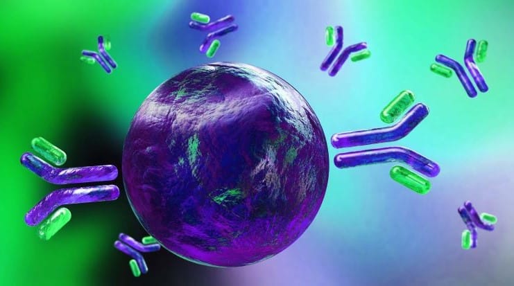
Flow cytometry is a technique for multi-parameter analysis of individual cells suspended in solution. It involves using fluorophore-labeled antibodies to detect specific cellular markers, and allows researchers to determine how many cells of a given type are present in the sample, if those cells express a particular protein, whether that protein is functionally active, and other critical insights.
A major advantage of flow cytometry over methods such as ELISA and western blot is that it provides information at the single-cell level. And, compared to microscopy-based techniques like immunocytochemistry (ICC) and immunohistochemistry (IHC), which offer this kind of detail, the higher throughput of flow cytometry produces data with greater statistical relevance.
When the first fluorescence-based flow cytometer reached the market in 1968, researchers were restricted to detecting just a handful of markers using antibodies they had labeled themselves. However, advances in instrumentation, software, and antibody-labeling technology mean that it is now possible to detect as many as 20 or more markers simultaneously at rates of over 10,000 cells/second.
Flow Cytometry Applications
Immunophenotyping, the process of detecting and quantifying different cell types within a mixed population, is one of the most common flow cytometry applications. Immunophenotyping has utility for studying normal growth and development, investigating the underlying mechanisms of disease, and developing novel drug candidates, as well as for many different diagnostic and prognostic applications.
Other flow cytometry applications include cell cycle analysis, monitoring cell health, and interrogating cell signaling pathways, such as through detecting a specific post-translational modification (PTM) or tracking the nuclear translocation of a protein. In addition, flow cytometry forms the basis of a similar, but distinct, technique known as fluorescence activated cell sorting (FACS), during which one or more cell populations of interest are isolated from the sample to allow for further downstream analysis.
How does a flow cytometer work?
Flow cytometers use a combination of fluidics, optics, and electronics to analyze individual cells in suspension. After being injected into the central core of the fluidics system, the cells are directed into single file by an outer sheath fluid (a process known as hydroelectric focusing) and flow past an interrogation point where one or more lasers are focused.
As each cell moves through the interrogation point, it scatters the laser light and, at the same time, any fluorophore-labeled antibodies that are bound to the cell fluoresce. The resultant signals are then guided by a series of dichroic mirrors and filters toward photomultiplier tubes (PMTs) for detection. When the light reaches a PMT, it generates a voltage pulse (known as an event) that is used in producing a graphical representation of the sample.
Flow cytometry light scatter and fluorescence measurements
Light scatter is recorded as both forward scatter (FSC) and side scatter (SSC) by detectors situated in front of or adjacent to the laser source, respectively. While FSC provides an indication of cell size, SSC correlates with granularity; used in combination, these measurements enable the classification of major cell sub-populations, as well as cellular debris.
More detailed characterization of each sub-population requires the use of fluorophore-labeled antibodies for detecting specific cellular markers. Theoretically, it is possible to measure as many as 40 different markers in the same experiment; however, it is more common for researchers to detect 5-10 markers simultaneously.
What is meant by flow cytometry panel design?
Panel design refers to the process of selecting fluorophore-labeled antibodies for a multiplexed flow cytometry experiment. Critically, as well as demonstrating specificity for targets of interest, antibodies should be labeled with fluorophores that are a) compatible with the flow cytometer’s lasers and detectors and b) are suitable for use together without fluorescence spillover producing false positive results.
It is recommended that bright fluorophores be paired with scarce targets, and vice versa, and that fluorophores are spread across as many lasers and detectors as possible. Where the number of available lasers is limited, tandem dyes provide opportunities for increased panel flexibility. Online spectra viewer and panel builder tools are widely used to guide panel design.
We provide a broad range of fluorophore-labeled antibodies, which includes FITC, PE, and Alexa Fluor® conjugates as well as antibodies labeled with various tandem dyes.
What’s involved in a typical flow cytometry experiment?
Once sample material such as cultured cells, blood, or excised tissue has been collected, a typical flow cytometry experiment begins with creating a single-cell suspension. Methods vary depending on the sample type, but general good practice guidelines include passing samples through a cell strainer to prevent clumping and diluting cells to an appropriate concentration to avoid missing events or extending run times. The protocol then diverges according to whether extracellular or intracellular targets are of interest. Where samples will be stained for cell surface markers, it is usual to block non-specific binding sites (and, often, to also block Fc receptors) before proceeding directly to immunostaining.
Staining for intracellular markers instead requires that cells be fixed and permeabilized prior to these steps to ensure antibodies can access their targets. If both extracellular and intracellular targets will be detected in the same experiment, staining for extracellular markers before detecting intracellular targets helps prevent damage to cell surface epitopes. After washing to remove any unbound antibody reagents, samples are introduced into the flow cytometer for processing and analysis.
How to choose antibodies for flow cytometry
As well as demonstrating specificity for targets of interest and being labeled with suitable fluorophores, antibodies must be sourced from appropriate host species if the immunostaining protocol will employ indirect detection. In this situation, all primary antibodies within the same multiplexed experiment must be derived from different host species and the secondary antibodies used for detection should be cross-adsorbed to avoid off-target binding. Primary antibodies should also be of a different host species to the sample material to circumvent the issue of secondary antibody binding to endogenous proteins.
Using secondary antibodies that recognize only a certain antibody isotype or subclass is a popular approach to increase the number of targets that can be indirectly detected. During assay development, antibody concentrations should be carefully optimized, and staining conditions identified to suit all of the antibodies that will be used in the same panel.
For over three decades, SouthernBiotech has offered primary and secondary antibodies that are quality tested by flow cytometry. Our extensive portfolio includes fluorophore-labeled mouse, rat, and human isotype- and subclass-specific secondary antibodies, in addition to numerous secondary antibodies that have been cross-adsorbed against multiple species.


